View our collection of functional GPCR assays for Gq, Gi, and Gs GPCRs to uncover agonists, allosteric modulators, and biased agonism
There are approximately 800 G protein-coupled receptors (GPCRs) in the human genome, making them the largest family of druggable targets. As surface receptors, GPCRs play roles in a variety of physiological functions including neurotransmission, sensation, immunity, and hormonal regulation, and they are the primary target for 36% of all approved drugs on the market.
Advances in our understanding of GPCR biology combined with novel screening assays to probe GPCR pharmacology have accelerated GPCR drug discovery. GPCR assays have led to the development of allosteric modulators, discovery of novel ligands and biased agonists, and repurposing of known drugs into new indications. At ION Biosciences, we leverage these tools with our state-of-the-art instrumentation to provide you with unparalleled insights for your GPCR discovery programs.
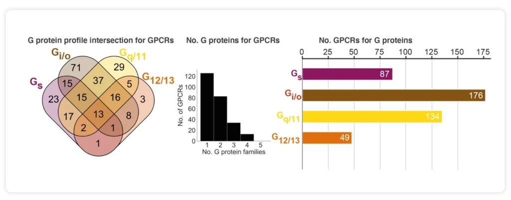
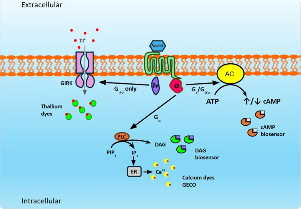
GPCRs have independent downstream signaling pathways that are determined by the alpha subunit of the G protein. The major alpha subunits are Gs, Gi/o, and Gq, and most GPCRs preferentially couple to one primary alpha subunit. However, many GPCRs can couple to multiple G proteins, and some couple directly to ion channels, inviting the exploration of pathway biased agonists. Gs and Gi GPCRs activate or inhibit adenylate cyclase, respectively, altering the levels of intracellular cAMP. Some Gi/o GPCRs also couple directly to GIRK channels, activating the channel upon agonist binding to promote potassium transport. Gq GPCRs measurable secondary messengers are calcium release from intracellular stores into the cytoplasm and the production of DAG from PIP2.
We quantify GPCR signaling dynamics using a collection of fluorescent ion indicators and biosensors to fully profile your compounds of interest. Furthermore, our unique fluorescent ion indicator portfolio and assays that we manufacture in house, alongside our expert team, can unlock even more benefits for your GPCR screening needs.
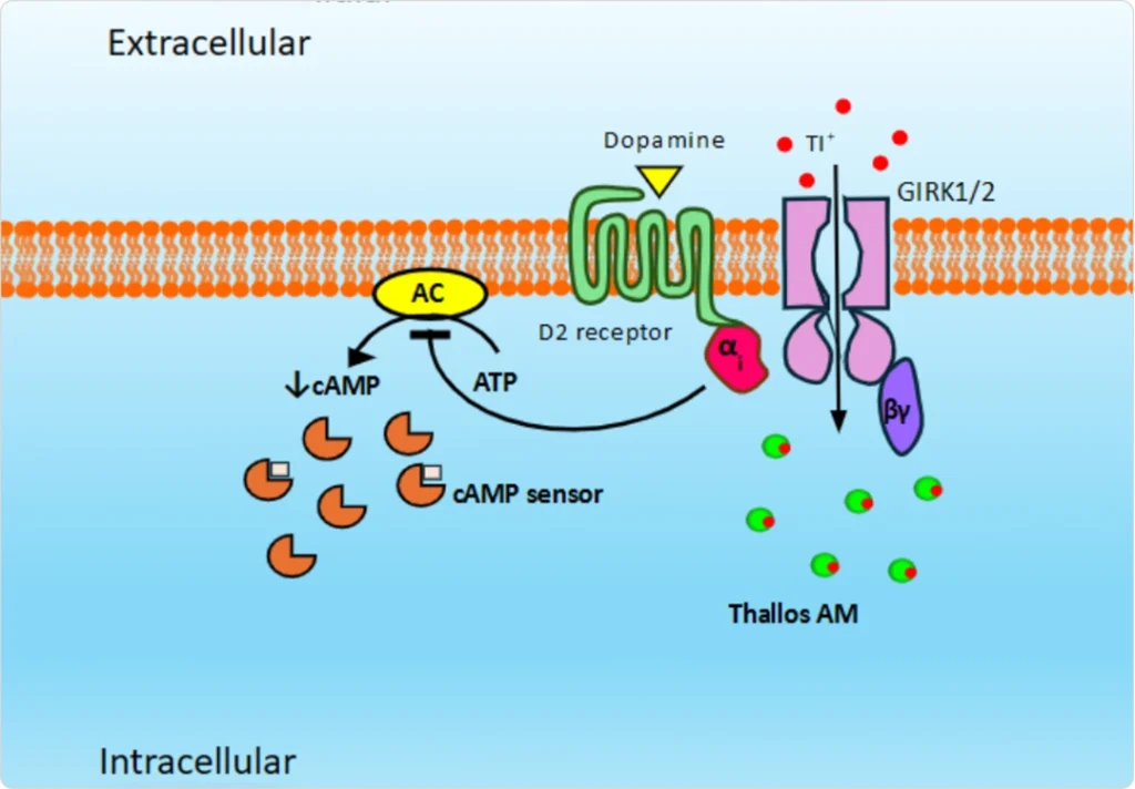
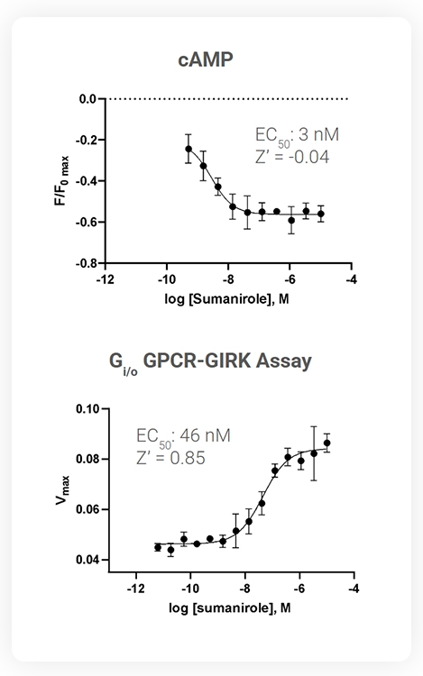
After agonist binding, the alpha subunit of Gi-coupled GPCRs inhibit adenylate cyclase (AC), resulting in a decrease in intracellular cAMP levels. Using cADDis, a fluorescent cAMP biosensor from Montana Molecular, and forskolin to artificially elevate cAMP levels, Gi GPCR agonism can be detected.
Another key signaling pathway for Gi/o-coupled GPCRs, is through activation of G protein-gated Inward Rectifying Potassium (K⁺) channels, better known as GIRK channels. Not only is the GPCR-mediated modulation of GIRK channels of great physiological importance, it also provides a convenient means of measuring Gi GPCR activity using fluorescence-based thallium flux assays.
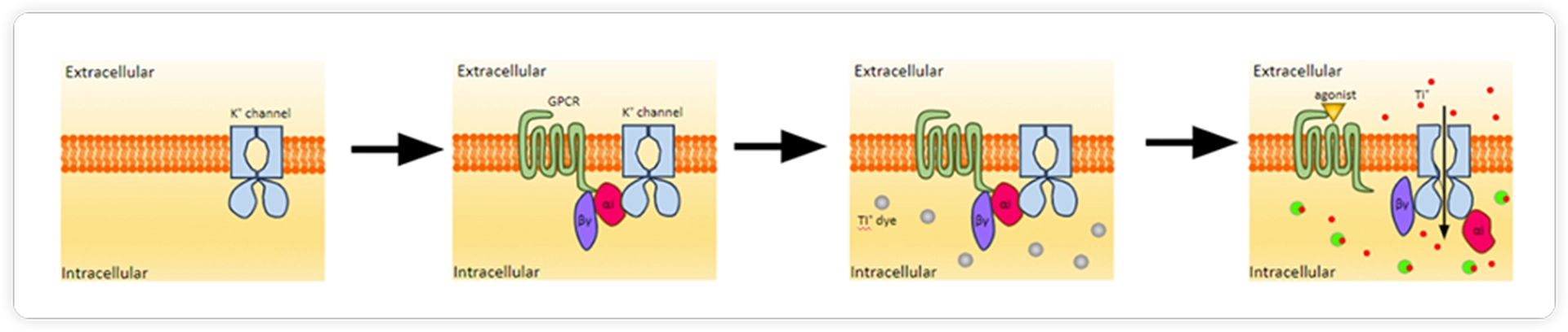
A GIRK-based Gi GPCR assay offers key advantages compared to other approaches that are used to discover modulators of Gi/o GPCRs:
Explore how our off-the-shelf solution can provide more details about the pharmacology of your Gi/o GPCR modulators.
The alpha subunit of Gi GPCRs inhibit adenylate cyclase (AC), resulting in a decrease in intracellular cAMP levels. A change in cAMP can be detected using cADDis, a fluorescent cAMP biosensor from Montana Molecular. For these assays, forskolin is used as a direct potentiator of AC to elevate intracellular cAMP levels, enabling visualization of AC inhibition upon ligand binding.
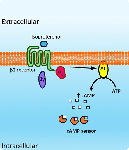
After agonist binding, Gs GPCRs activate adenylate cyclase (AC), an enzyme that converts ATP to cAMP. An increase in intracellular cAMP levels can be detected using cADDis, a fluorescent cAMP biosensor from Montana Molecular that provides a real-time readout of this important second messenger. Gs GPCR assays can be conducted in either endpoint or kinetic mode, where we can assess important ligand-dependent signaling dynamics.
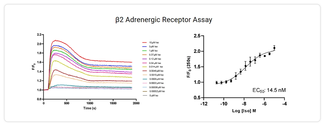
Kinetic profiles of cAMP generation triggered by stimulation of the 𝛃2 adrenergic receptor (ADRB2) with isoproterenol (Iso). ADRB2 is endogenously expressed in HEK293 cells, and cAMP dynamics are monitored in real-time using cADDis. A corresponding 14-pt dose response curve is also displayed.
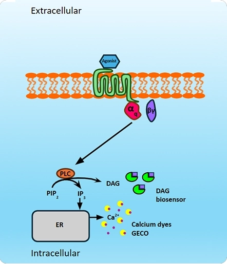
When Gq GPCRs are activated by ligands, they initiate a signaling cascade that involves the activation of phospholipase C-β (PLC-β) and subsequent production of diacylglycerol (DAG) and inositol trisphosphate (IP3). IP3 results in calcium release from intracellular stores into the cytoplasm via the IP3 receptor. Using fluorescent calcium assays, the IP3 signaling pathway has been employed as a rapid and effective means of identifying modulators of Gq GPCRs.
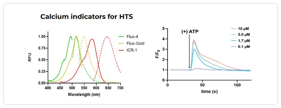
Fluorescence spectra of calcium indicators for HTS and a kinetic profile of a purinergic receptor assay. Our proprietary calcium indicators include Fluo-Gold, ICR-1, and BAPTA JF549, enabling multiplexing with the Panoptic HTS reader.
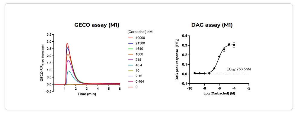
Biosensor assay examples for the M1 muscarinic receptor using GECO, a genetically encoded calcium indicator, and a DAG sensor from Montana Molecular.